Genes are the basic units of inheritance in nature. This article is the first in a four-part series exploring the role of genes and chromosomes in inheritance, health and disease
Abstract
Genes are passed down the generations in a predictable manner and we receive roughly half of our genetic material from each parent. This article explains the nature, structure and role of genes, deoxyribonucleic acid and chromosomes, describes how chromosomes determine gender, and touches on chromosomal abnormalities. It is the first article in a four-part series exploring the role of genes and chromosomes in inheritance, health and disease.
Citation: Knight J, Andrade M (2018) Genes and chromosomes 1: basic principles of genetics. Nursing Times [online]; 114: 7, 42-45.
Authors: John Knight and Maria Andrade, both senior lecturers in biomedical science at the College of Human Health and Science, Swansea University.
- This article has been double-blind peer reviewed
- Scroll down to read the article or download a print-friendly PDF here
- Click here to see other articles in this series
Introduction
With the exception of identical twins, all humans are genetically unique. During conception, a paternal sperm cell (spermatozoon) fuses with a maternal egg cell (ovum). The resulting embryo will have received approximately half of its genetic material from the father and half from the mother. Therefore, although all children are genetically unique, they typically exhibit a mix of characteristics inherited from both parents. The basic unit of inheritance in nature is the gene and genes are passed down the generations in a predictable manner. Each gene stores information in the form of deoxyribonucleic acid (DNA).
Cells and DNA
The average adult human body is composed of approximately 50 trillion (50 million million) cells. Most DNA is located in the nuclei of these cells; a much smaller amount is in the mitochondria – these are tiny bean-shaped organelles present in the cytoplasm of nucleated cells, which release the energy these need to survive.
The set of instructions – or genetic blueprint – used to construct the proteins that provide structure in all living organisms is encoded in the genes in the form of nucleic acids, the most common of which is DNA. This is true of the most simple viruses and bacteria all the way through to complex multicellular animals, including humans.
Composition and shape
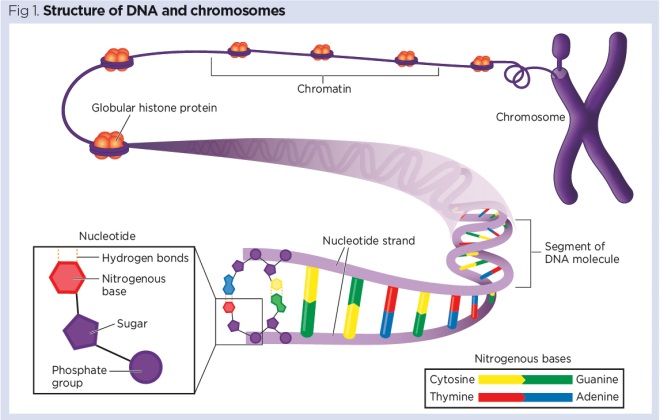
The building blocks of DNA are called nucleotides (VanPutte et al, 2017). Each nucleotide is constructed from a:
- Five-carbon sugar (deoxyribose);
- Phosphate group;
- Nitrogenous base.
In total, there are four nitrogenous bases, as follows:
- Adenine;
- Cytosine;
- Guanine;
- Thymine.
Molecules of DNA adopt the characteristic shape of a double helix. It is helpful to visualise them as a long ladder that has been twisted into a spiral shape, with the sugar and phosphate groups forming the rails and the nitrogenous bases forming the rungs (Fig 1). DNA has exceptional storage capacity (Box 1).

Box 1. DNA and bioinformatics
DNA can store information in a very stable and compact form. It has been calculated that all the data in the world (including every book, photograph and video) could be encoded into approximately 1kg of DNA (Extance, 2016). This has opened up a whole new area of research in the field of bioinformatics, which is currently exploring the use of DNA as a digital storage medium.
Complementary base pairing
The nitrogenous bases in DNA pair up in a predictable manner:
- Adenine always pairs with thymine (A-T);
- Cytosine always pairs with guanine (C-G).
This ‘complementary base pairing’ occurs because the bases structurally complement each other, fitting together like two pieces of a jigsaw puzzle. As a consequence, if the sequence of one strand of the DNA molecule is known, the other strand can be deduced (Box 2).
There are many online tools that use the base pairing rule to ascertain the complementary sequences for any given DNA sequence. Complementary base pairing also allows the genetic code to be easily replicated during cell division and translated into proteins.
Box 2. Deducing a complementary DNA sequence
If the sequence on one strand of DNA is ATCGATGCC, then the complementary sequence will necessarily be TAGCTACGG. This will give the following complementary base pairing sequence:
ATCGATGCC
TAGCTACGG
Purines and pyramidines
In addition to forming the base letters of the genetic code, adenine, cytosine, guanine and thymine are intimately involved in cell signalling, regulating enzyme activity and cellular metabolism. According to their chemical structure, the four nitrogenous bases are divided into two subgroups:
- Purines – this includes denine and guanine.
- Pyramidines – this includes cytosine and thymine.
Problems with metabolising purines can result in the common joint disorder, gout (Box 3).
Box 3. How gout develops
In later life, some people develop problems with metabolising purines (adenine and guanine), which can lead to a build-up of uric acid crystals in tissues and joints. This results in gout, which is a common disorder. As all foods are derived from plants or animals, which contain their own DNA, the human diet is naturally rich in purines. However, certain foods – notably offal, oily fish, shellfish and fermented grain products such as beer – are particularly rich in purine and can, therefore, exacerbate gout. Limiting the intake of purine-rich foods often relieves the symptoms of gout.
Mitochondrial DNA
Although most human genes are encoded in the DNA located in the nuclei of cells, mitochondria contain some of our DNA. Mitochondrial DNA (mtDNA) has 37 genes, many of which code for the production of enzymes involved in cellular metabolism (Chial and Craig, 2008). During fertilisation, spermatozoa usually only deliver nuclear DNA, so mtDNA is inherited solely along maternal lines via the ova.
Like bacteria, mitochondria can replicate by binary fission – a process that allows the mitochondrial genes to be passed on to ‘daughter’ mitochondria. Many of the genes carried by mitochondria are similar to those found in bacteria, which has led to the theory that mitochondria were once free-living bacteria that primitive cells incorporated into their cytoplasm.
DNA and chromosomes
The average human cell is around 20µm in diameter (a micron is one-thousandth of a millimetre). Most of a cell’s DNA is stored in its nucleus, which is even smaller: around 2-10µm in diameter (Milo and Phillips, 2016). Despite their small size, each nucleated human cell manages to pack in around 3m (10 feet) of DNA.
To make this possible, the DNA is neatly wrapped around histone proteins that function like a spool or bobbin. DNA and histone proteins form a material called chromatin, which is visible in the cell nucleus in the form of microscopic granules (VanPutte et al, 2017).
When a cell is preparing to divide, the DNA is wound up tighter and tighter and folds upon itself into coils. This tightly wound DNA is denser and thicker, and appears in the nucleus in the form of thread-like structures called chromosomes.
Chromosomes and gender
Human cells (except spermatozoa and ova) have 23 pairs of chromosomes, giving a total of 46 chromosomes; this is known as the diploid number. During cell division (mitosis), the diploid number is maintained. The chromosomes in the first 22 pairs – the autosomes – are the same in both sexes. The 23rd pair determines the gender of the individual; its two chromosomes are called the sex chromosomes (VanPutte et al, 2017). In most people, sex chromosomes come in one of two combinations:
- XX = female;
- XY = male.
As female cells only contain X chromosomes, ova will only ever contain X chromosomes. However, male cells always contain both X and Y chromosomes, so spermatozoa can have either an X or Y chromosome associated with them. Roughly equal numbers of X-bearing and Y-bearing spermatozoa are produced, so sex is determined by which type of spermatozoon fuses with the ovum. This normally results in roughly half of all children being male and half female (Fig 2).

There are only two possibilities when determining sex (X-bearing spermatozoon producing a girl or Y-bearing spermatozoon producing a boy) so it is a bit like flipping a coin – it is possible to see four or five heads (or more) in a row and, in the same way, parents may have four or five children (or more) of the same sex in a row. However, when populations are examined as a whole, there is roughly a 50:50 split between the sexes.
Not all males are XY and not all females are XX; indeed, some of the most common survivable chromosomal abnormalities affect the sex chromosomes. These will be discussed in part 4 in this series.
Karyographs and karyotyping
Photographs of chromosomes can be taken in actively dividing cells and then arranged into pairs according to size using special computer software. These photographs, known as karyographs, reveal an individual’s chromosomal make-up or karyotype.
Karyotyping is often carried out during pregnancy. In a procedure known as amniocentesis, a sample of the amniotic fluid (which surrounds the foetus in the amniotic sac) is taken using a needle. The amniotic fluid contains cells from the foetus, shed during intrauterine movement and when the fluid is breathed in and out of the foetal lungs. As the foetus is in a continual state of growth, most cells will be dividing and so chromosomes will be visible.
Karyotyping can reveal many things about an unborn child, including:
- Its gender;
- Whether the diploid number of chromosomes is present;
- Whether there are any extra chromosomes and which pair has the extra copy;
- Whether there are missing chromosomes and which pair has the missing copy;
- Whether a particular chromosome is too long;
- Whether a particular chromosome is too short.
Aneuploidy and Down’s syndrome
Sometimes karyotyping will reveal extra or missing chromosomes: such deviations from the diploid number are referred to as aneuploidy. The World Health Organization estimates that aneuploidy is present in at least 5% of pregnancies.
The most common aneuploidy is Down’s syndrome, or trisomy 21, in which people inherit an extra copy of chromosome 21. Each nucleated cell has 47, not 46, chromosomes. Having this extra copy results in affected individuals developing common physical and pathophysiological features (Mundakel and Lal, 2018), which include:
- Macroglossia (enlarged and often protruding tongue);
- Pronounced epicanthal folds (giving the eyes an oriental appearance);
- Learning difficulties and reduced intelligence quotient;
- Increased risk of heart defects, particularly septal defects;
- Increased risk of age-related diseases, including arthritis and dementia, earlier in life;
- Reduced lifespan.
Today, people tend to have children later in life and it is increasingly common for women to have their first child in their 40s. As a result, more children are born with chromosomal abnormalities such as Down’s syndrome. Karyotyping provides a useful prenatal screening tool, so parents, in conjunction with health professionals, can make informed decisions about the pregnancy and potentially prepare for the birth of children with specific medical or care needs. The fact that the age of the mother affects the likelihood of chromosomal abnormalities is linked to a phenomenon called non-disjunction, which will be discussed in part 4.
Gender selection
As karyotyping is a relatively simple procedure and can reveal the sex of the unborn child, it can be misused for gender selection. In most circumstances, it is illegal in the UK to terminate a pregnancy based on the sex of the foetus; it is only allowed in very exceptional cases based on serious medical grounds, such as one parent carrying a sex-linked genetic disease.
Chromosomes and genes
Genes are arranged linearly along the length of each chromosome (like beads on a string), with each gene having its own unique position or locus. In a pair of chromosomes, one chromosome is always inherited from the mother and one from the father. This means that, with the exception of genes on the sex chromosomes of males, we have two copies of each gene, one inherited from our mother and one from our father. These pairs of genes, which control many of our characteristics, are called alleles.
Genotype and phenotype
Each pair of genes that is inherited for a particular trait is referred to as the genotype for that trait; the physical characteristics that result from possessing those genes is referred to as the phenotype (VanPutte et al, 2017). It can be said that:
- The genotype is an individual’s genetic make-up;
- The phenotype is the physical appearance of the individual, which is determined largely by their genes but also affected by environmental factors.
Dominant and recessive genes
Some genes exert more influence than others: the former are called dominant and the latter are called recessive. For example, individuals may inherit genes for both blood group A and blood group O, in which case the genotype for that particular trait would be AO. In the case of blood groups, the A gene is dominant and the O gene is recessive, so an individual with the genotype AO for that trait would always have the phenotype blood group A. Dominant and recessive genes will be explored in more detail in part 4.
Structural and control genes
Genes can be divided into structural genes and control (or regulatory) genes.
Structural genes contain sequences of DNA coding for proteins such as:
- Actin and myosin (used to build muscle), keratin (found in skin, hair and nails) and haemoglobin (found in red blood cells);
- Enzymes driving the metabolic processes of the body;
- Hormones maintaining homoeostasis (for example, insulin);
- Neurotransmitters that help to co-ordinate physiological processes (for example, substance P).
Control or regulatory genes, as their name implies, control or regulate the activity of structural genes – including when these are switched on or off – and how much protein is produced. Control genes ensure that only certain genes are turned on in certain tissues; for example, the pancreas does not require the genes coding for keratin or haemoglobin to be switched on, but it is crucial to have the genes coding for insulin or glucagon switched on. Control genes are often themselves controlled, directly or indirectly, by other chemical signals including hormones, growth factors and neuro-transmitters.
The switching on and off of genes is also influenced by environmental factors including diet, exercise and pollutants. Epigenetics, a relatively new branch of genetics, examines the factors that can influence gene activity without actually changing any of the DNA sequences in the genome (Weinhold, 2006).
A revolution in the making
In 1990, the laborious process of sequencing the entire human genetic sequence, or genome, started. The human genome project ended ahead of schedule in 2003 and its results have been fine-tuned since then. In 2014 the human genome was thought to consist of 60,155 genes of which 19,881 were structural genes coding for proteins.
The original human genome project took over 12 years to complete and cost $2.7bn. Today, the entire genome of an individual can be sequenced in a few hours for around $1,000. That price is predicted to drop to around $100 in the next few years (Herper, 2017). Rapid genomic sequencing will likely revolutionise medicine in the coming decades and targeted therapies tailored to an individual’s genetic code could become commonplace.
Key points
- Genes store information in the form of deoxyribonucleic acid (DNA) located in the cells
- When a cell is preparing to divide, DNA takes the form of thread-like structures called chromosomes
- Most human cells have 23 pairs of chromosomes, giving a total of 46 – the diploid number
- Having extra or missing chromosomes is called aneuploidy, the most common condition of which is Down’s syndrome
- Structural genes, which contain sequences of DNA coding for proteins, are controlled by regulatory genes
Also in this series
Extance A (2016) How DNA could store all the world’s data. Nature; 537: 7618, 22-24
Herper M (2017) Illumina promises to sequence human genome for $100 – but not quite yet. Forbes; 9 January.
Milo R, Phillips R (2016) Cell Biology by the Numbers. Abingdon: Garland Science.
Mundakel GT, Lal P (2018) Down Syndrome. Medscape.
VanPutte CL et al (2017) Seeley’s Anatomy and Physiology. New York, NY: McGraw-Hill.
Weinhold B (2006) Epigenetics: the science of change. Environmental Health Perspectives; 114: 3, A160-A167.


Have your say
or a new account to join the discussion.