electron microscopy
- Key People:
- Richard Henderson
- Joachim Frank
- Jacques Dubochet
- Related Topics:
- microscope
- electron microscope
- cryo-electron microscopy
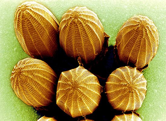
electron microscopy, Technique that allows examination of samples too small to be seen with a light microscope. Electron beams have much smaller wavelengths than visible light and hence higher resolving power. To make them more observable, samples may be coated with metal atoms. Because electrons cannot travel very far in air, the electron beam and the sample must be kept in a vacuum. Two different instruments are used. In the scanning electron microscope, a moving beam of electrons is scanned across a sample; electrons scattered by the object are focused by magnetic “lenses” to produce an image of the object’s surface similar to an image on a television screen. The images appear three-dimensional; they may be of small organisms or their parts, of molecules such as DNA, or even of large individual atoms (e.g., uranium, thorium). In the transmission electron microscope, the electron beam passes through a very thin, carefully prepared sample and is focused onto a screen or photographic plate to visualize the interior structure of such specimens as cells and tissues.


