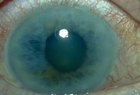
What are scleritis and sclera?

Scleritis is inflammation of the sclera, the white portion of the eye.
The sclera is the tough, white fibrous outer wall layer of the eyeball. It is a type of connective tissue. The sclera provides both the white color of the eye and maintains the shape of the eyeball. It extends from the edge of the cornea (the clear, dome-shaped window in the front of the eye) around to the optic nerve in the back of the eye.
What is necrotizing scleritis?
Necrotizing scleritis is the most severe and destructive form of scleritis, sometimes leading to loss of the eye from multiple complications, or occasionally perforation of the eyeball (globe). In necrotizing scleritis, there is a breakdown of the scleral tissue.
What is the sclera?
The sclera is the tough, white fibrous outer wall layer of the eyeball. It is a type of connective tissue. The sclera provides both the white color of the eye and maintains the shape of the eyeball. It extends from the edge of the cornea (the clear, dome-shaped window in the front of the eye) around to the optic nerve in the back of the eye.
Scleritis vs. episcleritis
Many of the conditions associated with scleritis are serious. Episcleritis is a much more common and milder inflammation of the tissue.
The extreme pain of scleritis helps to differentiate it from other common causes of redness of the eyes such as episcleritis, which is not as painful.
Scleritis vs. conjunctivitis
Many of the conditions associated with scleritis are serious. Conjunctivitis is a much more common cause of red eye. The extreme pain of scleritis helps to differentiate it from other common causes of redness of the eyes such as conjunctivitis, which can cause itching and burning but is not exceptionally painful. There is usually no discharge from the eye in scleritis while there is often a discharge with conjunctivitis.

IMAGES
Scleritis See a picture of eye diseases and conditions See ImagesWhat is the main cause of scleritis?
Scleritis is an uncommon disease and is differentiated from episcleritis, which is inflammation of the surface membrane covering the sclera and is a more common eye condition. In episcleritis, only the superficial tissue between the white of the eye (sclera) and the blood vessel-filled covering (conjunctiva) is inflamed.
Approximately one-half of scleritis cases are associated with underlying diseases that affect the body internally (systemic diseases). Connective tissue disorders, autoimmune diseases, and generalized vasculitic abnormalities may all first appear as scleritis or manifest themselves as scleritis at some other time during the underlying disease.
Scleritis may be seen in association with the following:
- systemic lupus erythematosus,
- arthritis,
- other types of inflammatory arthritis (ankylosing spondylitis,
- reactive arthritis,
- gouty arthritis,
- psoriatic arthritis,
- relapsing polychondritis),
- polyarteritis nodosa,
- mixed connective tissue disease,
- progressive systemic sclerosis (scleroderma),
- granulomatous polyangiitis,
- polymyositis,
- Sjögren's syndrome,
- giant cell arteritis,
- inflammatory bowel disease, and
- allergic angiitis.
Scleritis may be the initial manifestation of these underlying illnesses. Some of these conditions are organ-threatening and potentially lethal.
Scleritis can also be the result of an infectious process caused by the following:
- bacteria including pseudomonas,
- fungi,
- mycobacterium,
- viruses, or
- parasites.
- Trauma,
- chemical exposure, or
- postsurgical inflammation can also cause scleritis.
No cause is found in some cases of scleritis.
Scleritis may affect either one or both eyes. In patients with disease in both eyes, an underlying systemic cause is almost always found.
What are risk factors for scleritis?
The peak incidence of scleritis is in people aged 40-50 years old. Women are more commonly affected than men. The presence of known autoimmune or connective tissue disease markedly increases the risk of scleritis.
What are signs and symptoms of scleritis?
What are the early signs of scleritis?
- In scleritis, there is redness in the blood vessels adjacent to the sclera. This redness can have a bluish or violet tinge. With repeated episodes or long-standing scleritis, the sclera can thin and the underlying brown choroid may become visible through the residual sclera.
- Scleritis may be nodular with multiple round or oval elevated areas of the sclera.
- Scleritis may also be necrotizing, resulting in areas of thinning and softening of the normally fairly rigid sclera.
- Areas of the absence of redness may be due to death (necrosis) of inflamed blood vessels.
- Scleritis can be characterized as being located in the front or back of the eye (anterior or posterior), depending on the location of the disease upon examination.
- Visual acuity can be decreased if there is a secondary clouding of the cornea or the lens of the eye.
- Intraocular pressure can be increased from the congestion of the blood vessels involved in draining aqueous fluid from the eye.
- Intraocular pressure can also be decreased if the ciliary body, which lies deeper than the sclera, is secondarily involved by inflammation.
- Disturbances of eye movement can be seen since the extraocular muscles can become irritated when they insert into the sclera. This can cause double vision.
Symptoms of Scleritis
Symptoms of scleritis include:
- Pain
- Redness
- Tearing
- Light sensitivity (photophobia)
- Tenderness of the eye
- Decreased visual acuity
Pain is nearly always present and typically is severe and accompanied by tenderness of the eye to touch. The pain may be boring, and stabbing, and often awakens the patient from sleep. The extreme pain of scleritis helps to differentiate it from other common causes of redness of the eyes, such as conjunctivitis or episcleritis.
There is usually no discharge from the eye in scleritis.
The discoloration that is caused by the inflammation can have a bluish hue and can involve the entire white of the eye or be localized to only one area.
Decreased visual acuity may be caused by the extension of scleritis to the adjacent structures, leading to inflammation of the cornea (keratitis) or the colored portion of the front of the eye (uveitis), glaucoma, cataract, and abnormalities of the retina.
Is scleritis contagious?
Scleritis is usually not infectious and, therefore, is not contagious. Infectious scleritis occurs primarily in eyes that have had surgery or trauma.
What specialists diagnose and treat scleritis?
Ophthalmologists treat scleritis. An ophthalmologist is a medical doctor who has specialized in the diagnosis and treatment of eye disease. Your ophthalmologist may consult with an immunologist or rheumatologist to assist in your treatment depending on the particular situation.
How do healthcare professionals diagnose scleritis?
Scleritis is usually diagnosed by the history and the clinical findings on slit lamp examination by an ophthalmologist. The slit lamp is a special viewing instrument that eye specialists use to stabilize the head while magnifying and viewing the structures of the eye.
To determine the cause of the scleritis, blood tests including rheumatoid factor, antinuclear antibodies, antineutrophil cytoplasmic antibodies, human leukocyte antigen typing, and erythrocyte sedimentation rate may be ordered.
- If infectious disease is suspected, appropriate cultures or serological tests may be necessary.
- If granulomatosis with polyangiitis is considered, sinus X-rays and a chest X-ray may be ordered.
- Radiographic examination of joints may assist in the diagnosis of various types of arthritis.
- If orbital inflammation is suspected in addition to the scleritis, an MRI of the orbit may be helpful for diagnosis.
Subscribe to MedicineNet's Daily Health News Newsletter
By clicking Submit, I agree to the MedicineNet's Terms & Conditions & Privacy Policy and understand that I may opt out of MedicineNet's subscriptions at any time.
What is the treatment for scleritis?
Treatment of scleritis resulting from an underlying disease process usually requires specific therapy for that disease.
Topical treatment with eye drops is an adjunct to such systemic treatment. These eye drops will usually be anti-inflammatory, such as topical steroid drops or topical nonsteroidal anti-inflammatory drops (NSAIDs). Topical antibiotics are used if the scleritis is felt to be infectious.
In situations where no underlying disease process is found, eye drops to counter inflammation are used, but they are often insufficient to control the process. Systemic treatment with NSAIDs, cortisone medication (corticosteroids), or immune-modulating agents such as methotrexate (MTX) can be the first choice. But azathioprine, mycophenolate mofetil, cyclophosphamide, or cyclosporine are also used. Anti-TNF agents such as the biologics infliximab (Remicade) or adalimumab (Humira) can also be used.
Localized, subconjunctival steroid injections are often helpful in certain situations or if systemic side effects of these drugs are of concern.
The delivery of steroid eyedrops using a low-level electrical current to encourage drug migration into the affected tissue, known as iontophoresis, is currently in clinical trials.
Rarely, surgical procedures may be required if there is scleral thinning. To preserve the integrity of the eye, scleral grafts available through eye banks can be used. Corneal tissue may also be used if there is a perforation or severe thinning in the limbal area.
Are there home remedies for scleritis?
What is the prognosis for scleritis?
Scleritis is a serious eye disease that must be evaluated, treated, and monitored aggressively to avoid vision loss. Scleritis may be recurrent but is usually responsive to therapy. Any underlying disease must be diagnosed and treated. Many of the conditions associated with scleritis are serious and may only be diagnosed during the evaluation for the cause of the scleritis. The scleritis itself may respond to treatment, but the underlying disease process may not.
In general, scleritis in association with granulomatosis with polyangiitis is difficult to treat and may lead to vision loss even with treatment. The scleritis seen with spondyloarthropathies is usually very responsive to treatment. Scleritis that occurs in the absence of an underlying disease will typically respond well to treatment. When recurrent scleritis symptoms are noticed, urgent treatment can result in optimal outcomes.
What are the complications of scleritis?
Complications of scleritis include:
- Inflammation of the cornea (keratitis)
- Anterior or posterior uveitis
- Glaucoma
- Cataract
- Retinal swelling
- Scleral thinning
- Peripheral corneal shinning
- Retinal macular swelling
Corneal or scleral thinning, if untreated, may lead to a hole in the side of the eye (ocular perforation) and severe vision loss or blindness. Scleritis may be recurrent. Long-term treatment with corticosteroid eye drops may cause cataracts and glaucoma.
Is it possible to prevent scleritis?
Scleritis is an inflammation of the white of the eye. It is a serious eye disease that is often associated with underlying autoimmune disorders.
- Prompt diagnosis and treatment are essential in preventing permanent vision loss.
- There is no preventive treatment for most cases.
- Patients with underlying disease processes should be made aware of the possibility of scleritis occurring and should have access to immediate care and careful monitoring by an ophthalmologist.
What research is being done on scleritis?
Current research in scleritis is trying to determine the exact immune abnormalities leading to the disease and is searching for more precise medication to target those portions of the immune system.

QUESTION
The colored part of the eye that helps regulate the amount of light that enters is called the: See AnswerHodson, Kelly L., et al. "Epidemiology and Visual Outcomes in Patients With Infectious Scleritis." Cornea 32.4 April 2013: 466-472.
Johnson, Keegan S. and David S. Chu. "Evaluation of sub-Tenon triamcinolone acetonide injections in the treatment of scleritis." American Journal of Ophthalmology 149.1 (2010): 77–81.
Watson, Peter, et al. Duane's Ophthalmology, 15th Ed. Philadelphia, PA: Lippincott Williams & Wilkins
Zamir, Ehud, et al. "A prospective evaluation of subconjunctival injection of triamcinolone acetonide for resistant anterior scleritis." Ophthalmology 109.4: 798-805
Top Scleritis Related Articles

Blindness
Blindness is the state of being sightless. Causes of blindness include macular degeneration, stroke, cataracts, glaucoma, infection, and trauma. Symptoms and signs may include eye pain, eye discharge, or the cornea or pupil turning white. Treatment of blindness depends upon the cause of the blindness.
Chest X-Ray
Chest X-Ray is a type of X-Ray commonly used to detect abnormalities in the lungs. A chest X-ray can also detect some abnormalities in the heart, aorta, and the bones of the thoracic area. A chest X-ray can be used to define abnormalities of the lungs such as excessive fluid (fluid overload or pulmonary edema), fluid around the lung (pleural effusion), pneumonia, bronchitis, asthma, cysts, and cancers.
Common Eye Problems
Eye diseases can cause damage and blindness if not treated soon enough. Learn the warning signs and symptoms of common eye conditions such as glaucoma, cataracts, pink eye, macular degeneration and more.
Eye Picture
The eye has a number of components which include but are not limited to the cornea, iris, pupil, lens, retina, macula, optic nerve, choroid and vitreous. See a picture of the Eye and learn more about the health topic.
Eye Conditions Quiz
What do you know about your eyes? Take this quick quiz to learn about a range of eye diseases and conditions.
Inflammatory Bowel Disease
The inflammatory bowel diseases (IBD) are Crohn's disease (CD) and ulcerative colitis (UC). The intestinal complications of Crohn's disease and ulcerative colitis differ because of the characteristically dissimilar behaviors of the intestinal inflammation in these two diseases.
MRI (Magnetic Resonance Imaging Scan)
MRI (or magnetic resonance imaging) scan is a radiology technique which uses magnetism, radio waves, and a computer to produce images of body structures. MRI scanning is painless and does not involve X-ray radiation. Patients with heart pacemakers, metal implants, or metal chips or clips in or around the eyes cannot be scanned with MRI because of the effect of the magnet.
Pinkeye
Pinkeye, also called conjunctivitis, is redness or irritation of the conjunctivae, the membranes on the inner part of the eyelids, and the membranes covering the whites of the eyes. These membranes react to a wide range of bacteria, viruses, allergy-provoking agents, irritants, and toxic agents.
Pink Eye Slideshow
How do you get pink eye? And how contagious is pinkeye? If you woke up with crusty eyelids and red, swollen eyes, you may have conjunctivitis. Learn about eye drops and home remedies for pink eye.
Rheumatoid Arthritis (RA)
Rheumatoid arthritis (RA) is an autoimmune disease that causes chronic inflammation of the joints, the tissue around the joints, as well as other organs in the body.
Rheumatoid Factor
Rheumatoid factor is often measured in blood tests for the diagnosis of rheumatoid arthritis. However, rheumatoid factor can also be present in individuals with other conditions such as lupus, infectious hepatitis, syphilis, mononucleosis, tuberculosis, liver disease, and sarcoidosis.
Erythrocyte Sedimentation Rate
Erythrocyte sedimentation rate is a common blood test that is used to detect and monitor inflammation in the body. It is performed by measuring the rate at which red blood cells (RBCs) settle in a test tube. The erythrocyte sedimentation rate is simply how far the top of the RBC layer has fallen in one hour, increasing with more inflammation.
Lupus (Systemic Lupus Erythematosus or SLE)
Lupus is a condition characterized by chronic inflammation of body tissues caused by autoimmune disease. Lupus can cause disease of the skin, heart, lungs, kidneys, joints, and nervous system. When internal organs are involved, the condition is called systemic lupus erythematosus (SLE). When only the skin is involved, the condition is called discoid lupus.
What Are the Causes of a Headache Behind the Eyes?
A headache behind the eyes is an uncomfortable sensation that is felt around or on the back of the eye, which may or may not be a throbbing ache. Causes of headaches behind the eyes include tension headaches, migraines, cluster headaches, sinus headaches, occipital neuralgia, brain aneurysm, Grave's disease, scleritis, dry eyes, vision problems, eye strain and poor posture.
